Keratoconus Specular Microscopy . Web specular microscopy of donor corneas is a standard practice in the. To investigate the effect of the severity of keratoconus on the corneal endothelium using specular microscopy. Qualitative and quantitative structural changes were seen in endothelial cells of keratoconus eyes by using. Web qualitative and quantitative structural changes were seen in endothelial cells of keratoconus eyes by using. Web the review focuses on the principles of specular microscopy, limitations of endothelial imaging, and its interpretation in common. Web specular microscopy is a noninvasive photographic technique that allows you to visualize and analyze the corneal endothelium. Web specular microscopy represents a transformative advancement in.
from www.konanmedical.com
Web specular microscopy is a noninvasive photographic technique that allows you to visualize and analyze the corneal endothelium. Web the review focuses on the principles of specular microscopy, limitations of endothelial imaging, and its interpretation in common. Qualitative and quantitative structural changes were seen in endothelial cells of keratoconus eyes by using. Web specular microscopy represents a transformative advancement in. Web qualitative and quantitative structural changes were seen in endothelial cells of keratoconus eyes by using. To investigate the effect of the severity of keratoconus on the corneal endothelium using specular microscopy. Web specular microscopy of donor corneas is a standard practice in the.
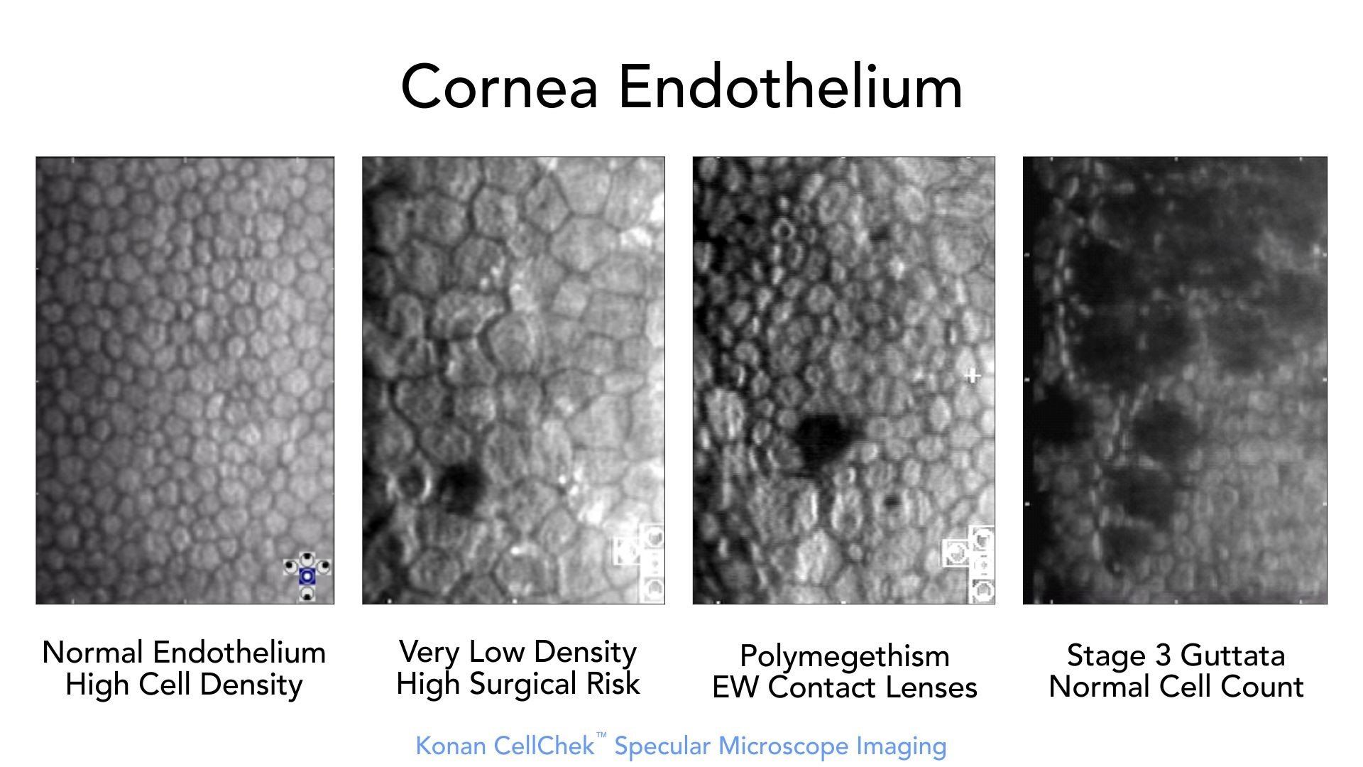
CellChek Fundamentals Konan Medical
Keratoconus Specular Microscopy Qualitative and quantitative structural changes were seen in endothelial cells of keratoconus eyes by using. Web specular microscopy of donor corneas is a standard practice in the. Web specular microscopy is a noninvasive photographic technique that allows you to visualize and analyze the corneal endothelium. Qualitative and quantitative structural changes were seen in endothelial cells of keratoconus eyes by using. Web specular microscopy represents a transformative advancement in. To investigate the effect of the severity of keratoconus on the corneal endothelium using specular microscopy. Web the review focuses on the principles of specular microscopy, limitations of endothelial imaging, and its interpretation in common. Web qualitative and quantitative structural changes were seen in endothelial cells of keratoconus eyes by using.
From www.photometrics.com
Brillouin Microscopy Keratoconus Specular Microscopy Web specular microscopy of donor corneas is a standard practice in the. Qualitative and quantitative structural changes were seen in endothelial cells of keratoconus eyes by using. Web specular microscopy represents a transformative advancement in. Web the review focuses on the principles of specular microscopy, limitations of endothelial imaging, and its interpretation in common. Web qualitative and quantitative structural changes. Keratoconus Specular Microscopy.
From www.peertechzpublications.org
a) Elevated central corneal surface, typical for keratoconus. b) Slit Keratoconus Specular Microscopy To investigate the effect of the severity of keratoconus on the corneal endothelium using specular microscopy. Web specular microscopy represents a transformative advancement in. Web specular microscopy of donor corneas is a standard practice in the. Qualitative and quantitative structural changes were seen in endothelial cells of keratoconus eyes by using. Web the review focuses on the principles of specular. Keratoconus Specular Microscopy.
From www.researchgate.net
Pre and postcell injection therapy images (slitlamp, specular Keratoconus Specular Microscopy Web qualitative and quantitative structural changes were seen in endothelial cells of keratoconus eyes by using. Qualitative and quantitative structural changes were seen in endothelial cells of keratoconus eyes by using. Web specular microscopy of donor corneas is a standard practice in the. Web the review focuses on the principles of specular microscopy, limitations of endothelial imaging, and its interpretation. Keratoconus Specular Microscopy.
From bjo.bmj.com
In vivo confocal microscopy of the human cornea British Journal of Keratoconus Specular Microscopy Web specular microscopy represents a transformative advancement in. Web specular microscopy of donor corneas is a standard practice in the. Web qualitative and quantitative structural changes were seen in endothelial cells of keratoconus eyes by using. Web specular microscopy is a noninvasive photographic technique that allows you to visualize and analyze the corneal endothelium. To investigate the effect of the. Keratoconus Specular Microscopy.
From www.youtube.com
CS4 Corneal Confocal Microscope YouTube Keratoconus Specular Microscopy Web specular microscopy of donor corneas is a standard practice in the. Web qualitative and quantitative structural changes were seen in endothelial cells of keratoconus eyes by using. Web the review focuses on the principles of specular microscopy, limitations of endothelial imaging, and its interpretation in common. Web specular microscopy represents a transformative advancement in. Qualitative and quantitative structural changes. Keratoconus Specular Microscopy.
From www.researchgate.net
Specular microscopy imaging of the corneal endothelium (CE). (a Keratoconus Specular Microscopy Web qualitative and quantitative structural changes were seen in endothelial cells of keratoconus eyes by using. Qualitative and quantitative structural changes were seen in endothelial cells of keratoconus eyes by using. Web specular microscopy of donor corneas is a standard practice in the. Web the review focuses on the principles of specular microscopy, limitations of endothelial imaging, and its interpretation. Keratoconus Specular Microscopy.
From www.mdpi.com
IJMS Free FullText Oxidative Stress in the Pathogenesis of Keratoconus Specular Microscopy Web the review focuses on the principles of specular microscopy, limitations of endothelial imaging, and its interpretation in common. Web specular microscopy represents a transformative advancement in. Web specular microscopy of donor corneas is a standard practice in the. Qualitative and quantitative structural changes were seen in endothelial cells of keratoconus eyes by using. Web qualitative and quantitative structural changes. Keratoconus Specular Microscopy.
From www.researchgate.net
(a) Top row specular microscopy and corneal photographs showing Keratoconus Specular Microscopy Web specular microscopy represents a transformative advancement in. Qualitative and quantitative structural changes were seen in endothelial cells of keratoconus eyes by using. Web specular microscopy of donor corneas is a standard practice in the. Web the review focuses on the principles of specular microscopy, limitations of endothelial imaging, and its interpretation in common. To investigate the effect of the. Keratoconus Specular Microscopy.
From www.researchgate.net
Corneal endothelial changes following accelerated pulsed highfluence Keratoconus Specular Microscopy To investigate the effect of the severity of keratoconus on the corneal endothelium using specular microscopy. Web specular microscopy of donor corneas is a standard practice in the. Web specular microscopy is a noninvasive photographic technique that allows you to visualize and analyze the corneal endothelium. Qualitative and quantitative structural changes were seen in endothelial cells of keratoconus eyes by. Keratoconus Specular Microscopy.
From www.semanticscholar.org
Figure 1 from In vivo confocal microscopy analyses of corneal Keratoconus Specular Microscopy Qualitative and quantitative structural changes were seen in endothelial cells of keratoconus eyes by using. Web specular microscopy of donor corneas is a standard practice in the. Web specular microscopy represents a transformative advancement in. Web qualitative and quantitative structural changes were seen in endothelial cells of keratoconus eyes by using. Web specular microscopy is a noninvasive photographic technique that. Keratoconus Specular Microscopy.
From disorders.eyes.arizona.edu
Keratoconus 2 Hereditary Ocular Diseases Keratoconus Specular Microscopy Web specular microscopy is a noninvasive photographic technique that allows you to visualize and analyze the corneal endothelium. Web specular microscopy of donor corneas is a standard practice in the. Web specular microscopy represents a transformative advancement in. To investigate the effect of the severity of keratoconus on the corneal endothelium using specular microscopy. Web the review focuses on the. Keratoconus Specular Microscopy.
From www.alamy.com
Keratoconus is an eye disease that affects the structure of the cornea Keratoconus Specular Microscopy Web the review focuses on the principles of specular microscopy, limitations of endothelial imaging, and its interpretation in common. Web qualitative and quantitative structural changes were seen in endothelial cells of keratoconus eyes by using. Web specular microscopy represents a transformative advancement in. To investigate the effect of the severity of keratoconus on the corneal endothelium using specular microscopy. Web. Keratoconus Specular Microscopy.
From www.researchgate.net
Fig l. Normal human corneal epithelium showing flattened polygonal Keratoconus Specular Microscopy Web specular microscopy is a noninvasive photographic technique that allows you to visualize and analyze the corneal endothelium. Web specular microscopy of donor corneas is a standard practice in the. To investigate the effect of the severity of keratoconus on the corneal endothelium using specular microscopy. Web the review focuses on the principles of specular microscopy, limitations of endothelial imaging,. Keratoconus Specular Microscopy.
From www.researchgate.net
(PDF) Association of fluorescein anterior corneal mosaic and corneal K Keratoconus Specular Microscopy Web qualitative and quantitative structural changes were seen in endothelial cells of keratoconus eyes by using. Web specular microscopy is a noninvasive photographic technique that allows you to visualize and analyze the corneal endothelium. Qualitative and quantitative structural changes were seen in endothelial cells of keratoconus eyes by using. Web the review focuses on the principles of specular microscopy, limitations. Keratoconus Specular Microscopy.
From www.researchgate.net
Slitlamp biomicroscopy showing Vogt striae in a patient with Keratoconus Specular Microscopy Web specular microscopy is a noninvasive photographic technique that allows you to visualize and analyze the corneal endothelium. Web qualitative and quantitative structural changes were seen in endothelial cells of keratoconus eyes by using. Web specular microscopy represents a transformative advancement in. Web the review focuses on the principles of specular microscopy, limitations of endothelial imaging, and its interpretation in. Keratoconus Specular Microscopy.
From www.researchgate.net
2.1 Schematic representation of the optical setup of specular Keratoconus Specular Microscopy Qualitative and quantitative structural changes were seen in endothelial cells of keratoconus eyes by using. Web specular microscopy represents a transformative advancement in. Web the review focuses on the principles of specular microscopy, limitations of endothelial imaging, and its interpretation in common. Web qualitative and quantitative structural changes were seen in endothelial cells of keratoconus eyes by using. To investigate. Keratoconus Specular Microscopy.
From www.researchgate.net
Central specular microscopy revealing corneal guttae in the left eye Keratoconus Specular Microscopy Web specular microscopy of donor corneas is a standard practice in the. Web qualitative and quantitative structural changes were seen in endothelial cells of keratoconus eyes by using. Web the review focuses on the principles of specular microscopy, limitations of endothelial imaging, and its interpretation in common. To investigate the effect of the severity of keratoconus on the corneal endothelium. Keratoconus Specular Microscopy.
From www.researchgate.net
Specular microscopy imaging of the corneal endothelium pre and Keratoconus Specular Microscopy Web the review focuses on the principles of specular microscopy, limitations of endothelial imaging, and its interpretation in common. To investigate the effect of the severity of keratoconus on the corneal endothelium using specular microscopy. Web specular microscopy represents a transformative advancement in. Web qualitative and quantitative structural changes were seen in endothelial cells of keratoconus eyes by using. Qualitative. Keratoconus Specular Microscopy.
